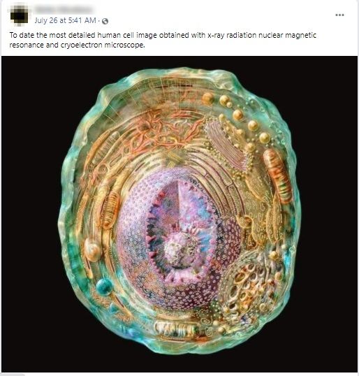
A photo is being shared on social media claiming it as the detailed human cell picture obtained with x-ray radiation, nuclear magnetic resonance, and cryo-electron microscope. Let’s verify the claim made in the post.

Claim: Photo of the detailed human cell picture obtained with x-ray radiation, nuclear magnetic resonance, and cryo-electron microscope.
Fact: This photo shows an illustrated picture of an animal cell, not the detailed image of human cell. This illustrated animal cell poster was designed by an Australia-based science illustrator, Russel Kightley. Hence, the claim made in the post is FALSE.
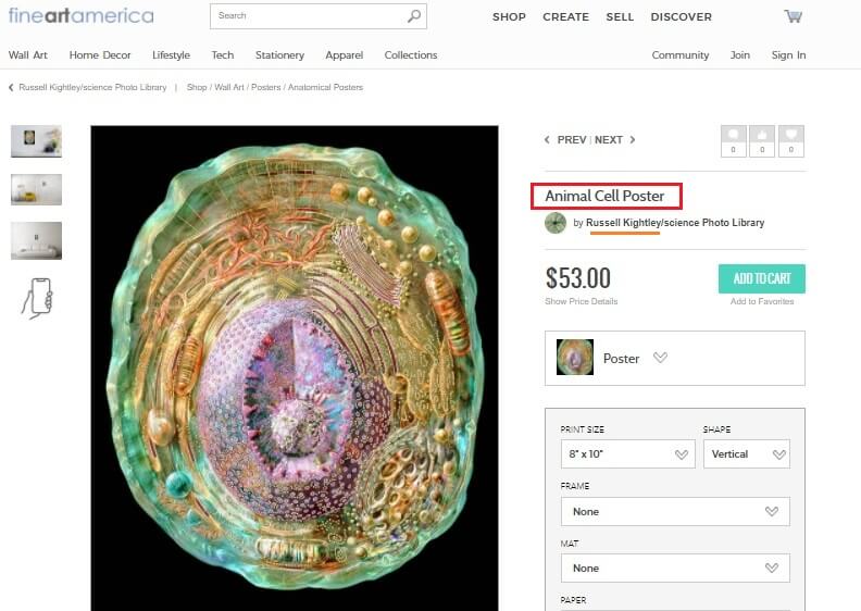
On reverse image search of the photo shared in the post, a similar photo is found on the ‘Fine Art America’ website. This website mentioned it as an animal cell poster designed by Russel Kightley. The ‘Pixels’ and ‘Imgur’ image-sharing websites have also shared the same image with a similar description.

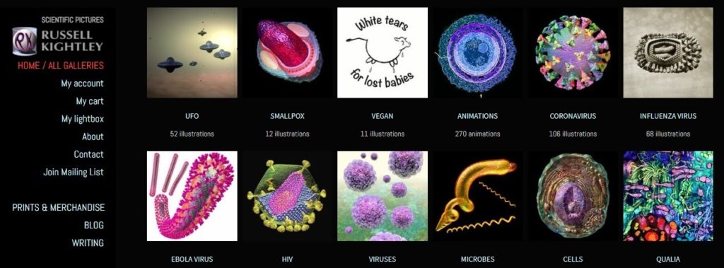
Russel Kightley, an Australia-based science illustrator had created this animal cell image and shared it in his portfolio. Russel Kightley is working as a science illustrator since 1981.

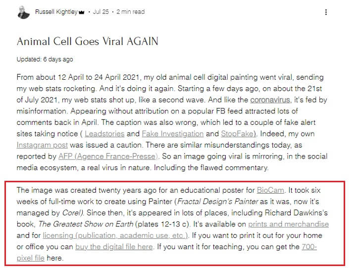
When his illustrated animal cell image has recently gone viral as the human cell image, Russel Kightley issued a clarification on his website. Russel Kightley said, “The image was created twenty years ago for an educational poster for BioCam. It took six weeks of full-time work to create using Painter (Fractal Design’s Painter as it was, now it’s managed by Corel). Since then, it’s appeared in lots of places, including Richard Dawkins’s book, The Greatest Show on Earth (plates 12-13 c)”. He also issued a clarification on Instagram.

From all these pieces of evidence, it can be concluded that the image shared in the post shows an illustrated animal cell picture, not the detailed image of a human cell.
To sum it up, an illustrated animal cell picture was falsely shared as the detailed image of a human cell.


